A Seven-year Sickness? Tracking Sindbis virus in Swedish Mosquitoes

From: “Sindbis virus polyarthritis outbreak signalled by virus prevalence in the mosquito vectors”, J.O. Lundström, J.C. Hesson, M.L. Schäfer, Ö. Östman, T. Semmler, M. Bekaert, M. Weidmann, Å. Lundkvist and M. Pfeffer (2019), PLoS Neglected Tropical Diseases

https://doi.org/10.1371/journal.pntd.0007702

Sindbus virus (SINV) is a virus found across the world. Usually, it is transmitted between birds such as Fieldfare and Redwing (both of which are winter visitors to the UK) by mosquitoes. However, it sometimes makes the jump to humans (zoonosis) where it produces a disease characterised by joint pain and rashes, known variously as Pogosta disease (in Finland), Ockelbo disease (in Sweden) and Karelian Fever (in Russia).* Interestingly, it appears that SINV is mainly transmitted between birds by two almost indistinguishable species of mosquito Culex torrentium and Culex pipiens, but usually transmitted to humans by another species, Aedes cinereus. It has been suggested that SINV outbreaks occur on a seven-year cycle, but it is not easy to be sure, as records in humans seem to be fairly heavily influenced by how much interest governments were taking in the disease at the time. If outbreaks could be accurately predicted, the public could be forewarned to take protective measures. Even simple measures like using insect repellent and wearing more covering clothing could save doctors a lot of time and money, and patients a lot of pain.

Transmission of Sindbis Virus, taken from Lundström et al 2019

Jan Lundström’s team had a theory. They tested thousands of mosquitoes caught in light traps around eight lakes in central Sweden every other week from 2001 to 2003 for the presence of SINV, suspecting that an outbreak in mosquitoes may precede an outbreak in humans.** They were also able to take genetic samples of the virus, in order to determine its outbreak history.

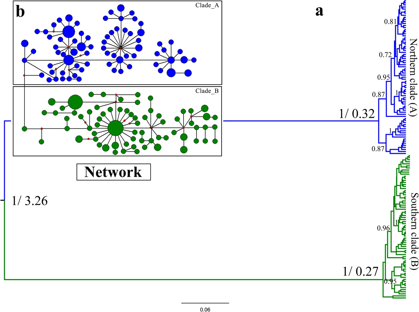
What they found was surprising. It seems that SINV is fairly new to Scandinavia, having arrived only a century or so ago, and slowly emerging into significance in the late 1960s and 70s. What’s more, it appears to have, somehow, arrived four separate times. The scientists don’t speculate on how this could have happened, but further research may yet turn up some very interesting finds.

The authors, reasonably, focus a lot more on how and when SINV turns up in their mosquitoes, both Aedes and Culex. On the basis of the seven-year theory, they thought that 2002 would be an outbreak year, and, happily, it was found that only a small proportion of mosquitoes in 2001 were infected, rising sharply in 2002 and then declining again in 2003. Intriguingly, all the 2001 virus samples were found in Culex mosquitoes, but it then appeared in Aedes cinereus in 2002, allowing it to make the jump to humans, and was only found in Aedes in 2003. This might not mean anything, but it does hint at the possibility that SINV has to reach a certain prevalence in birds before Aedes cinereus will pick it up and transmit it to humans. When the scientists looked at mosquitoes in 2009, seven years after 2002, they again found an increase in the prevalence of SINV, suggesting that spikes in the number of mosquitoes infected with SINV coincide with outbreaks in humans. Which makes perfect sense, when the mosquitoes are the things transmitting the virus.

It has been noticed before that in Sweden, SINV cases appear in front of doctors from July to October, most commonly in mid to late August. The researchers found that the mosquitoes followed the same pattern. This study detected SINV in mosquitoes between the 30th July and the 10th September, and a previous one found a slightly earlier, but very similar time period of the 16th July to the 30th August. Not only does this give Swedes a timeframe to be particularly wary of mosquito bites, it also gives researchers looking for SINV in mosquitoes a tighter window, with less time wasted looking for the virus when it is less likely to be around. This means that it will be a lot easier to properly test the seven-year outbreak theory, using mosquitoes as a more reliable record of how prevalent SINV is. Human medical records are great, but you can’t assume that every person infected is going to show up to a doctor’s surgery or that no records have been lost. It could be that many more people than we think get infected, but never become ill enough to seek treatment. With mosquitoes, you can have a good stab and finding out exactly what percentage of the population has the virus, and track that fairly easily and accurately over time, with a far simpler database, and far fewer other factors to worry about. Mosquitoes live much simpler lives than we do – you don’t have to worry about whether their diet or exercise habits are skewing your data. The authors also suggest an effort to sample mosquitoes in early July could pay off in giving doctors a warning signal of when SINV cases in humans are likely to appear, and the public a warning of when to be careful. This, again, would save a lot of time and trouble.

It seems that it’s still early days for research into Sindbis virus, but I for one look forward to seeing how well the seven-year theory stands up to further testing.

*For more information on Sindbus virus see the European Centre for Disease Prevention and Control here: https://ecdc.europa.eu/en/sindbis-fever/facts

** I don’t know why this is only getting published nearly 20 years later, but this sort of thing does happen in science sometimes, for all kinds of reasons

If you find any inaccuracy or lack of clarity in this article, please don’t hesitate to contact me on j.d.r1612@gmail.com and I’ll fix it as soon as I am able

Is it better to be different? How water fleas resist parasites

From: “Daphnia parasite dynamics across multiple Caullerya epidemics indicate selection against common parasite genotypes”, E. González-Tortuero, J. Rusek, P. Turkode, A. Petrusek, I. Maayan, L. Piálek, C. Tellenbach, S. Gießlerc, P. Spaak, J. Wolinska (2019), Zoology 119:4

https://doi.org/10.1016/j.zool.2016.04.003

Parasite-host interactions are rapidly becoming a major frontier in evolutionary biology, and are increasingly recognised a major driver of genetic, physiological and behavioural diversity. After all, two species locked into a constant arms race on which their survival depends are under pretty serious pressure to evolve something new. Many studies into host-parasite ‘coevolution’ have been carried out on the Daphnia water flea and its various parasites. Daphnia, microscopic crustaceans enjoying a blameless existence filtering algae from fresh water,are an excellent model for studying evolution because they have a short life cycle (almost always under a year), are easy to keep, have a variety of parasites,  and tend to live in very separate populations in different ponds. This allows for comparisons between different ponds, and a way to test if any trends in evolution are consistent between different populations of the same species. Daphnia even have a resting (ephippia) stage which can sit, dormant, in the mud for many years. Some very exciting research has been done on reviving resting Daphnia from many years ago, and seeing how differently they respond to the parasites of today than modern Daphnia.

Enrique González-Tortuero and colleagues were investigating Daphnia from a Swiss lake called Greifensee. What makes this lake special for evolutionary biologists is that the Daphnia in it have been tested for a parasite called Caullerya mesnili biweekly in the summer and monthly in the winter (life is pretty slow in alpine lakes in the winter) since 2002. From 2007, these tests also sampled bits of their DNA, and their parasite’s DNA. The scientists had a theory that a phenomenon known as Negative Frequency-Dependant Selection was in play in Greifensee, and set out to test this.

Negative Frequency-Dependant Selection (NFDS) is, like many terms in biology, a mouthful describing a fairly simple concept. The idea is that in a dangerous world, it is beneficial to be different. If a parasite has evolved to be very good at targeting a specific feature, any host that has an unusual version of it that gives the parasite trouble is going to be at an advantage. Because of this advantage, their descendants flourish, until they become so common that the parasites evolve a way round their defence. Those parasites with a way round the defences of this now-common host type flourish, and become more common. When this happens, it now pays to be different to this type, and the original type may become more common as the new type decreases.

Imagine a left-handed tennis player. Most of their opponents will be used to playing right-handers, and will be confused by the left-hander’s movements. This gives our leftie an advantage. Now imagine that left-handed players became so common they’re a majority, and every right-hander has played them many times. Our leftie has had their advantage nullified, and it is rapidly becoming advantageous to be right-handed. If right-handers then become a majority again, left-handers are back at an advantage. And so on and so on.

These situations do occur in nature, particularly if a parasite has evolved a specialist way to attack a feature controlled by a single gene or set of genes. Say a deadly virus latches onto a particularly shaped membrane protein to help it invade a cell (not an uncommon event at all). Individuals with an even slightly differently shaped protein are going to be better at resisting the virus – until they become so common that the virus adapts to the different shape. Now individuals with the other shape may be less vulnerable to the virus, and become more common.

But is this happening to the Daphnia in Greifensee? Quite possibly. The researchers found that between 2010 and 2013 the most common genetic type (genotype) of the C. mesnili parasite, known at CAUL-1, which was almost ubiquitous in 2010 has become less common over time. Around 80% of C. mesnili had it in 2010, falling to 60% by 2013.* At the same time, vanishingly rare (only detected once in a sample) genotypes proliferated. Likewise, there were many rare Daphnia genotypes and a few common ones, suggesting there is something advantageous about having a rare genotype, and that no single genotype is by itself advantageous enough to make up the entire population, as would be expected in such isolated populations if there were no parasites.

Proportions of different C. meslini genotypes in Greifensee over 4 years. Taken from González-Tortuero et al (2019)

At first sight, this seems like evidence for NFDS, suggesting that the Daphnia resistant to whatever it is that CAUL-1 does becoming common enough for CAUL-1 be far less advantageous.  However, the authors note that they cannot rule out an environmental change pressuring against individuals with CAUL-1 (they don’t know of any environmental changes, but they can’t prove there’ve been none). Neither can they rule out the possibility that some other event, perhaps a mass die-off, has affected the genetics of the parasites. Furthermore, NFDS requires that both the host and the parasite are genetically specific to each other (as they are genetically adapting to each other), but the evidence seems to be against that in these Daphnia and C. meslini, on the basis of the author’s tests.

The evidence is ambiguous, as it often is in real science, but it is possible that for these Daphnia and Caullerya meslini in Greifensee, it is better to be different.

*It’s not quite as simple as I’m making out there. 60% of the genotypes detected in 2013 were CAUL-1, but the DNA is being extracted from bits leftover inside the Daphnia, which matches up well with the individuals in the population of C. mesnili, but isn’t quite the same thing.

If you find an error or a lack of clarity in this piece, please don’t hesitate to contact me on j.d.r1612@gmail.com and I’ll fix it as soon as I am able

A House Divided: what splits a lineage of birds?

From “Deep south-north genetic divergence in Godlewski’s bunting (Emberiza godlewskii) related to uplift of the Qinghai-Tibet Plateau and habitat preferences”, J. Li, G, Song, N. Liu, Y. Change and X. Bao (2019), BMC Evolutionary Biology 19:161

From almost the beginning of recorded history through to the twentieth century the basic unit of Chinese politics has been the dynasty, lineages of powerful men and women who built and warred and reformed over successive generations. And while human families were branching off, moving about, and changing over the centuries, so were those of other species. Godlewski’s bunting (Emberiza godlewskii) is a small, brown, grey and orangey-pink bird, not dissimilar to a linnet, found across a large area of Asia. Like any animal with such a large range, it exhibits its fair share of genetic and visible (phenotypic) variation over its range.

Jiande Li and colleagues sought to investigate this variation by extracting DNA from 190 blood samples taken from 26 sites across China (the buntings were released immediately after donating blood). What they found was not totally unexpected, but surprising enough for those of us who aren’t experts in Chinese fauna: there are two, very distinct groups of Godlewski’s bunting, one from the north of China, and one from the south. The authors calculate that these two lineages split off from one another between 2.53 and 4.08 million years ago, with their best estimate of the date of this divergence being 3.26 million years ago. During these millions of years of separation, the two lineages seem to have hardly interacted at all: the scientists calculate that in a single generation, on average, 0.091 individuals migrated from the northern population to the southern one, and 0.077 went the other way. That is, a northern individual arrived in the south only once every 11 generations, and a southern individual travelled north every 13 generations, on average. By examining patterns in the DNA of the buntings, the researchers were also able to make a few more deductions, such as that the northern population had expanded rapidly 0.05 million years ago, while the southern one expanded in a much more modest fashion 0.12 million years ago. They also found that the family tree (phylogram) they produced from the bunting’s DNA had several subgroups in both the north and the south, but these didn’t quite match up to the subspecies that are currently recognised. This just goes to show how cladistics (organising organisms by their common ancestors) doesn’t always march hand-in-hand with traditional taxonomy (classifying organisms by visible traits). This doesn’t necessarily make traditional taxonomy wrong – it is quite easy to get slightly different answers out of most cladistics methods depending on which bits of information about the organism you put in, and common ancestors are not always the most useful way of grouping organisms to a naturalist in the field anyway. That said, biology has largely adopted cladistics as the future, and we are rapidly approaching the point where we have enough computing power to process many organisms’ entire DNA data all at once to build ever more accurate family trees.

The ‘family tree’ of Godlewski’s buntings (Emberiza godlewskii ) in China. Lengths of lines are genetic difference, and numbers on the right indicate time, in millions of years, since the two groups at each junction diverged

But I’m getting sidetracked. The accuracy of cladistics at a small scale notwithstanding, it is pretty clear that our northern and southern buntings went their separate ways a couple of million years ago and have had very little contact since. How did this happen? The scientists propose that one of the main driving forces was the Qinghai-Tibet Plateau being lifted up by tectonic forces, creating a very different set of habitats. This also created valleys and basins that could have formed a refuge where the buntings could retreat during colder glacial periods. During this time, the movement of the plateau and climatic changes also moved parts of the Yunnan-Guizhou Plateau into the influence of the monsoon, creating ideal habitat for the Godlewski’s buntings in the south. Crucially, the buntings found themselves split on either side of the Quinling Mountains, which, being pulled up with the Quinghai-Tibet Plateau, also created more basins for the buntings to settle in. In the past million years, both northern and southern buntings took the opportunity created by the last warm interglacial period to expand their ranges further into China, as suitable habitat emerged.

So, to summarise – it appears that geological and climatic changes split a perfectly normal population of birds in two, causing them to go their genetically separate ways. It only makes you wonder what the genetic consequences of the current climate emergency are going to be.

If you find an error or a lack of clarity in this piece, please don’t hesitate to contact me on j.d.r1612@gmail.com and I’ll fix it as soon as I am able

Public health in termite cities: how insects prevent epidemics

From “Subterranean Termite Social Alarm and Hygienic Responses to Fungal Pathogens”, M.S. Bulmer, B.A. Franco and E.G. Fields (2019), Insects 10:8, 240

https://doi.org/10.3390/insects10080240

Throughout history, one of the biggest dangers of city living has been contagious disease. Where people live close to one another, germs and pathogens can easily pass between them, causing epidemics. From the Black Death*in the 14th Century to Cholera in the 19th** and Tuberculosis today, living in large, static groups brings huge dangers in addition to its many benefits. The same problems also face other social animals, such as termites. Like us, termites build huge cities, and like us, their lives are threatened by disease. And, like us, they have behaviours which control the spread of disease.

Mark Bulmer, Bruno Franco and Edith Fields decided to investigate these behaviours by exposing the underground-dwelling termites Reticulitermes flavipes to the pathogenic (disease-causing) fungus Metarhizium in a variety of ways. Some individuals or groups were exposed directly to different strains of the fungus in water (a Metarhizium suspension; different concentrations were also tested), and some were presented with a dead termite killed by Metarhizium. The researchers were looking mainly for two crucial behaviours: allogrooming, where the termites cleaned the fungus off each other, killing it with antibiotics*** in their saliva or else eating it (to be digested in their stomachs); and rapid bursts of longitudinal oscillatory movement, or LOMs, which indicates that the termite is alarmed.

As could be expected from such a complex experiment, they had a variety of results, not all of which are easy to interpret. Interestingly, several of these results indicate that individual termites vary in their responses to, and even in their ability to detect, different strains of Metarhizium. It also appears that LOMs, the alarm response of termites to the fungus, is a social phenomenon. Individual termites encountering Metarhizium suspension never displayed LOMs, but groups always did. Pairs did some of the time, seemingly due to the individual differences between termites. However, the first termites of a group of 12 to find a dead termite felled by Metarhizium were also the first in the group to show alarm. The researchers believe that this extra level of alarm shown by LOMs triggered the termites to group together and clean each other (allogrooming). This response appears to be effective: the higher the level of alarm shown by the termites, the lower their risk of death was. LOMs does not appear to affect the amount of antibiotics the termites produce, suggesting that the social response of allogrooming was more important in protecting the termites than the way their immune systems were ‘primed’ by the fungus. Different strains of Metarhizium also elicited different levels of alarm from the termites, and, in a very interesting twist, this appears to go some of the way to explaining differences in virulence and dangerousness of the different strains.

So: a social response to a pathogen, triggering termites to clean each other, seems to be more important in determining whether or not they are killed by disease than their individual immune systems. There’s probably a metaphor in there, if you like such things.

*Which, admittedly, did not in any way spare rural areas, burning through the countryside as quickly as it did the cities

**Yes, I know, my Eurocentric education is showing here

***technically, antimicrobials and antifungals, not antibiotics, but I’ll excuse myself the use of a more familiar term

If you find an error or a lack of clarity in this piece, please don’t hesitate to contact me on j.d.r1612@gmail.com and I’ll fix it as soon as I am able

Live fast, die young? Where personality is linked to altitude

From “Among-population divergence in personality is linked to altitude in plateau pikas (Ochotona curzoniae)”, J. Qu, D. Réale, Q.E. Fletcher and Y Zhang. (2019), Frontiers in Zoology 16:26

Plateau Pikas (Ochotona curzoniae) are rather cute rabbit-like animals which can be found across the Tibetan Plateu, north of India and on the western edges of China. Life history traits are any traits which can affect how long and in what manner an organism lives: rates of growth, timing of reproduction, whether it reproduces once or several times, how long it takes for the body to give into aging, or how aggressive it is, for example. Jiapeng Qu and colleagues, having found out that different life history traits can often be found in the same species at different altitudes (heights above sea level), set out to find out if this is true for personality in pikas. They already knew that pikas higher up tend to live more slowly, with a shorter breeding season, as well as smaller and fewer litters each year than those lower down, and had a particular reason for examining personality.

The Pace-of-Life Syndrome (or POLS) hypothesis suggests that life history traits which are involved in trade-offs between present and future can be placed along a slow-fast sliding scale. That is, organisms which live fast, die young, grow fast and mature quickly will also be less docile, more active and more aggressive. In principle, this makes perfect sense for two reasons – if you’re not going to live for longer than a year, you have far less to lose by any given risk than if you can expect to be alive in five years. On the other hand, if food is scarce, it is more beneficial to live slowly and produce offspring more slowly but also to be more docile in order to spend more time looking for food and less running away from potential threats. If food is plentiful, it costs you less to spend energy running from threats, and it is far easier to produce many offspring than if food is scarce. Essentially, ‘slower’ organisms prioritise future growth and reproduction, whereas ‘faster’ ones focus more on the present, breeding quicker, producing more offspring, moving about more, and being more aggressive.

Aiming to test this hypothesis, the authors of this paper, knowing that pikas at higher altitudes tend to live slower lives, decided to see how this affected their personalities. They looked at three entirely separate groups of pikas at three different altitudes, and used two tests of personality to assess docility (how long a pika remained still in a mesh bag) and activity (how far a pika moved around a square metre box). Having first tested whether individual pikas were consistent in their behaviour, they then analysed the results.

Their results did indeed provide some support the Pace-of-Life Syndrome hypothesis: the pikas from the higher altitudes were more docile and less active than those from medium altitudes, which were more docile and less active than those from lower altitudes. Generally speaking, more docile individual pikas were also less active, but this did not appear to be the case in the lowest altitude group. The differences between the groups are very likely to be due to different environments at different heights: for example, at higher altitudes, pikas which are more docile will spend less time running from potential dangers, perhaps allowing them more time to feed in a place where food is harder to come by. Lower down, it may be that the risks of being eaten outweigh the gains from the extra feeding time, as food is more plentiful. It may also be that pikas living on more open ground higher up, where there is less vegetation, are harder for predators to spot if they sit stiller. Pikas living lower down, where there is more vegetation to hide under, may be less visible when they move. The researchers suggest that this may be why docility and activity seem not to be linked in the lower groups of pikas – if it makes less difference whether a pika is docile or active, individuals can be both at once, moving around a lot, but running from danger less easily.

All in all, this is a clever little experiment, which shows how important the environment animals live in is to how they evolve to behave.

If you find an error or a lack of clarity in this piece, please don’t hesitate to contact me on j.d.r1612@gmail.com and I’ll fix it as soon as I am able

How melanic was my squirrel: how dark squirrels got their colour

From: “Multiple origins of melanism in two species of North American tree squirrel (Sciurus)”, H.R. McRobie, N.D. Moncrief and N.I. Mundy (2019), BMC Evolutionary Biology 19:140

https://doi.org/10.1186/s12862-019-1471-7

Melanin is one of the most common pigments in nature. It determines our hair and skin colour, as well as that of many other animals – the more melanin in your hair, the darker it is, and people with less melanin are blonder. In other animals, it is likewise very common for different individuals to have different melanin levels, and therefore different coat colours. Many of these colour variations across a whole host of species, from birds to mice, have been found to be associated with two genes, ASIP and MC1R.

Helen McRobie and her colleagues were interested to find out whether melanism (darker colouration resulting from higher production of melanin than is usual for a species) had evolved separately in two species of squirrel, and whether it was produced by the same process in both. They therefore examined the genetics of Eastern Fox Squirrels (Sciurus niger) and Eastern Grey Squirrels (Sciurus carolinensis: the ones you can find in any British park). Grey Squirrels look more or less the same everywhere, but Fox Squirrels in the north and west of their American range tend to be orange agouti, whereas the ones in the south-east are more silver-grey or tan agouti with black heads, and white noses and ears. Oddly, the scientists found that different genes appeared to be responsible for darker, melanic colouring in these two different colour forms of fox squirrel: the melanic orange agouti forms seemed to be due to a variant of MC1R, whereas the south-eastern grey/tan agouti forms seemed to develop melanic colouring due to a mutation in ASIP. This suggests rather strongly that melanism has evolved more than once in the same species. If something evolves independently more than once, that suggests that it is advantageous in certain circumstances. It is not entirely clear what advantages darker colouring gives a squirrel, but melanism can provide camouflage, or help an animal keep warm (darker surfaces absorb heat better). For the Grey Squirrels and the Fox Squirrels which live in the colder parts of America, the increased heat absorption from a dark coat may be useful. It has been noticed since the 1740s that a greater proportion of Grey Squirrels were melanic in the northern part of their range. But why would melanism occur in warmer places? It has been suggested that darker colouration provides more camouflage in areas frequently burned by wildfires (where there will be lots of burnt wood, charcoal and ash). An attempt to test this theory with hawks found that darker squirrels were harder to spot when moving, but the standard, lighter colour patterns seemed to be better camouflage when the squirrel was still. It is still something of a mystery why melanism evolves, but it seems to have done so twice in fox squirrels.

As for the origin of melanism in grey squirrels, the researchers found something even stranger. The mutated form of MC1R which is associated with melanism in Grey Squirrels is known as MC1R∆24, as it is missing 24 base pairs (the building block of DNA). The exact same mutation is associated with melanism in the orange agouti Fox Squirrels. Moreover, the genes around it are the same. When genes are shuffled around during the production of sperm and egg, some genes stick together, and so tend to get inherited together. In the Grey Squirrel, there is a cluster of genes inherited with MC1R∆24 which look remarkable like fox squirrel genes. This appears to be because they are. It seems that Grey and Fox Squirrels sometimes interbreed, and once, when that happened, a hybrid Grey/Fox Squirrel inherited MC1R∆24, bred with a Grey Squirrel, and became the ancestor of the melanic Grey Squirrels that are around today. McRobie and her colleagues speculate that this hybrid squirrel may have had an advantage in colonising habitat different to where it’s ancestors live: perhaps the colder northern part of the Grey Squirrel’s range, where its darker coat helped it keep warm.

So, how did the squirrels become melanic? It seems very likely that two different mutations caused darker coats in Fox Squirrels, and one of these spread into Grey Squirrels. Which just goes to show, evolution has a knack for the unexpected.

If you find an error or a lack of clarity in this piece, please don’t hesitate to contact me on j.d.r1612@gmail.com and I’ll fix it as soon as I am able

Creating a growing cell: synthetic biology and the role of FtsZ in cell division

From: “Synthetic cell division via membrane-transforming molecular assemblies”, S. Kretschmer, K.A. Ganzinger, H.G. Franquelim and P. Schwille (2019), BMC Biology 17:43

https://doi.org/10.1186/s12915-019-0665-1

Cell membranes are the structures that make life possible. They are what keeps a patch of water having all the interesting biologically-important molecules* at high enough concentrations for all the chemical reactions which make up life to occur. As cell structures go, it is incredibly simple – a double layer of molecules that naturally arranges itself into a barrier. The membrane is made up of phospholipids, bizarre, but simple, accidents of chemistry. Each one is made up of a phosphate head, which is attracted to water, and a lipid tail, which repels water. This operates by exactly the same process by which oil poured into water will sullenly refuse to mix and form its own oily bubbles. When phospholipids are place in water, the phosphate heads move towards the water, and the lipid tails towards the other lipids, resulting in a double layer of lipid tails sandwiched between phosphate heads. This naturally curves round and forms a bubble. Stick in some DNA, amino acids, and a few other random bits and pieces, and you’ve very nearly got a cell. The cell membrane is not made up entirely of phospholipids, however – though some molecules (notably water and dissolved oxygen) can move through the membrane without difficulty, many cannot. The same membrane that keeps the biologically useful amino acids, DNA, ions, sugars and everything else in also keeps them out, and cells tend to die if they cannot access bring food into the cell and regulate concentrations of the chemicals inside them. For this reason, the cell membrane is also packed with molecule transporters, as well as receptors, external enzymes and such.

However, a cell can only get so big. As a cell grows, it must, inevitably, split off into two daughter cells. In single-celled organisms, the daughter cells go off to live independently, whereas in multicellular (many-celled) organisms like ourselves, the daughter cells continue to cooperate closely. (Colonial unicellular organisms like to blur that line, but that’s another story). For an organism to grow, its cells must divide. This involves splitting a single membrane, a single phospholipid bilayer, into two totally separate ones. Which brings us, in a roundabout way, to synthetic biology.

One of synthetic biology’s major aims, according to Simon Kretschmer and his colleagues, is to build a ‘minimal cell’. This minimal cell would, in theory, be designed from scratch, and do all the same basic processes a living cell does, most importantly taking in and processing energy from outside itself to maintain a stable environment inside itself. This would not only provide new avenues to explore the fundamentals of cell biology, what life is and how it first appeared, these minimal cells would also be incredibly useful bioreactors. Instead of having chemists manufacture important drugs, the machinery to make them biologically could be inserted into a minimal cell, which would then produce the drugs quite happily and very cheaply (an alternative, to do the same with existing bacteria, is already being explored). This is not quite as daft as it sounds: penicillin, the first modern antibiotic, is produced naturally by several species of Penicillium fungi, which was originally its sole source.

This minimal cell, if produced, would need to be able to divide. To do this, it would need to be able to split its cell membrane. As I mentioned earlier, all living things have molecular mechanisms to do this, which tend to be fairly consistent within kingdoms, but vary significantly between, for example, bacteria and animals.** Interestingly, these mechanisms are not entirely essential for cell division: bacteria without them (and also without cell walls) can still divide works by producing extra membrane and changing the shape of the membrane. Mycoplasma genitalium can even literally pull itself apart by moving each end in opposite directions if it’s on a solid surface. Synthetic membranes have even been found to be able to be divided by puffs of air, without losing anything inside them. It has been speculated that something like this may even have occurred on the early Earth. Despite these oddities, synthetic biologists have recently been focusing a lot of their efforts on a bacterial protein called FtsZ.***

FtsZ is rather strange. When a bacterial cell is ready to divide, FtsZ forms a ‘Z-ring’ of moving, overlapping filaments at the site where the cell is going to split. It anchors onto the cell membrane using two further proteins, FtsA and ZipA. Together these attract even more chemicals involved in cell division. It is not quite clear to what extent the forces that pull the cell apart are produced by FtsZ’s ‘treadmilling’, rather than the cell wall, but FtsZ does appear to play a role.

In synthetic ‘cells’, several studies have been able to do away with the anchors FtsA and ZipA by using a ‘fusion protein’, FtsZ-YFP-MTS, made from bits of FtsZ and a couple of other proteins. Not only is FtsZ-YFP-MTS able to deform membranes, under the right conditions it can also self-organise into rings. What is particularly intriguing is that the shape FtsZ-YFP-MTS seems to be largely determined by the concentration of magnesium ions around it, something which is fairly easily manipulated. Other synthetic biology experiments have been able to use only FtsZ with ZipA or FtsA to deform membranes. Crucially, the deformations produced by FtsZ, FtsZ-YFP-MTS and similar proteins have been suggested to be important in constriction and separating of the artificial membranes studied – i.e. they may contribute towards making the artificial ‘cell’ divide. However, it has still not been proven that FtsZ can produce enough force to split a membrane from the inside. Time will tell if FtsZ ends up being used to produce a minimal cell, but here and now it is certainly demonstrating the fundamental strangeness and complexity of cell biology.

*That is to say, Biochemists and Molecular Biologists claim that they are interesting. This has yet to be independently verified.

**This has some interesting implications for their evolutionary history – perhaps the different mechanisms arose independently?

***This is only one of the many areas of study covered by Kretschmer and colleagues, and my decision to focus on it is purely based on how good a story I can tell about it

If you find an error or a lack of clarity in this piece, please don’t hesitate to contact me on j.d.r1612@gmail.com and I’ll fix it as soon as I am able

Looking for Leishmaniasis in Bats and Dogs

From “Potential animal reservoirs (dogs and bats) of human visceral leishmaniasis due to Leishmania infantum in French Guiana”, H. Medkour, B. Davoust, F. Dulieu, L. Maurizi, T. Lamour, J-L. Marié, and O. Mediannikov (2019), PLoS Neglected Tropical Diseases 13:6

https://doi.org/10.1371/journal.pntd.0007456

Leishmaniasis is a neglected tropical disease caused by several species of the animal-like single-celled parasite Leishmania. It can infect many mammals, dogs being a particularly important, and is spread between hosts by sandflies. It is a versatile parasite, and though most of its hosts do not display any symptoms, dangerous infection can occur in both the skin (cutaneous leishmaniasis) and internal organs (visceral leishmaniasis). For this reason, it is something of an iceberg disease: almost half of infected dogs show no signs, for example, making it very hard to know just how common it is. As a variety of mammals, including bats, wild and domestic dogs, rodents and hares, are known to carry Leishmania, Hacène Medkour and colleagues decided to look for infections in bats and dogs in French Guiana.

French Guiana is a patch of France (technically an overseas department) in the Amazon, bordering Brazil to the south and Suriname to the west. Because of this, the French military frequently take working dogs on exercises in Guiana, which come from European France and go back after four months. This makes them potential carriers of Leishmania across continents. The scientists therefore tested three groups of animals for Leishmania: military working dogs, domestic dogs, and wild bats. Bats are a surprisingly common source of human diseases, particularly viruses (most notoriously SARS). As bats can also fly long distances, they can be important in transporting diseases to new places.

The researchers therefore took blood samples from 179 Guianese domestic dogs, 257 military dogs and 92 bats over ten years. They tested this blood for Leishmania DNA and RNA (a chemical very similar to DNA, which can be used to indicate where a particular gene is active). If the blood was tested positive for DNA or RNA, the sample was further tested for antibodies against Leishmania. The presence of these antibodies indicates that the host’s immune system is either fighting back against infection, or has cleared a Leishmania infection at some point in the past. Because only animals which already tested positive for Leishmania DNA were tested for the antibodies, past infection could be ruled out: the infection would definitely have been acquired recently, if the DNA was still present. Tests were performed both for generic Leishmania, and the species Leishmania infantum, which had previously been found in a dog in French Guiana.

Leishmania infantum was found in 3 (1.7% of) Guianese dogs, all in 2013. Between 2012 and 2018, 7 (9.0% of) military dogs were found to have acquired Leishmania in Guiana, 2 (2.6%) of which developed leishmaniasis. Only a single Seba’s short-tailed bat (1.1% of the studied bats; Carollia perspicillata) was found to have Leishmania, but it did not show any obvious symptoms of leishmaniasis. The authors suggest this is because there are so many large mammals in Guiana that the sandflies which transmit Leishmania have much more attractive targets than bats!

Though only a small number of the animals were found to have Leishmania, this is still significant, as Leishmania infantum is barely known in dogs in French Guiana: the last reported case was in a dog from Spain, in 2006. In humans, meanwhile, an average of 180 cases of leishmaniasis a year are reported in French Guiana, though none of these are due to Leishmania infantum. It is hard to know what to make of these results, but the researchers suggest that there may be a local transmission cycle for Leishmania in French Guiana. The fact that no domestic Guianese dogs in the study were infected before 2013 suggests that Leishmania infantum may be spreading, perhaps brought into Cayenne (the capital) from the rainforests where the military dogs frequently work. This, however, is a theory which will need testing more thoroughly. Like all good epidemiology papers, this one ends with a call for more active surveillance of the disease, in this case of the nonhuman animals which are potential sources of leishmaniasis. It just goes to show how much we still have to learn about where diseases are found and how they are transmitted, particularly in the tropics. They’re not called neglected tropical diseases for nothing.

For more information on leishmaniasis: https://www.who.int/leishmaniasis/en/

If you find an error or a lack of clarity in this piece, please don’t hesitate to contact me on j.d.r1612@gmail.com and I’ll fix it as soon as I am able

How do you put a number on what we haven’t discovered?

From: “Hidden in plain sight: what remains to be discovered in the eukaryotic proteome?” by V. Wood, A. Lock, M.A. Harris, K. Rutherford, J. Bähler and S.G. Oliver (2019), in Open Biology 9:2

https://doi.org/10.1098/rsob.180241

Proteins are the main movers in biochemistry. Genes code for proteins, and when the gene is expressed (activated), it produces its own particular protein. This protein can have almost any function, from digesting food to influencing the expression of other genes. Despite this, our knowledge of proteins remains incomplete, and for every protein that is well-understood by biologists, there are others that are not so well-known, or even completely unknown. To try to understand more about what remains to be discovered, Valerie Wood and her colleagues compared knowledge across three well-studied species: humans, and two yeasts, Saccharomyces cerevisiae (a ‘budding yeast’) and Saccharomyces pombe (a ‘fission yeast’). Yeasts are a common ‘model’ used to study biochemistry, because they are easy to find, easy to grow, easy to keep and surprisingly similar to humans, plants and animals.

The authors of this paper classified knowledge of proteins into three categories, using what is known as a ‘Gene Ontology’ approach:

  1. molecular function (MF) – where it is known what role the protein has in the molecular machinery
  2. biological process (BP) – where the role of the molecular system the protein is part of is known
  3. cellular component (CC) – where the location of a protein within a cell is known

In humans, out of 19,737 proteins, all three aspects were known for only 15,162 proteins. Interestingly, in 1,382 human proteins, none of the three aspects were known. The scientists also used another measure, based partly on distinguishing between proteins which were marked as ‘unknown’ in databases and those with no descriptions of their role at all. From this they estimated that across both yeasts and humans, around 20% (1 in 5) proteins have not been described in a useful way. Many of these proteins are found both in yeasts and in humans, suggesting that they have been maintained over millions of years of evolution. This implies that they are very important.

Knowledge of proteins in fission yeast (S. pombe), budding yeast (S. cerevisiae) and humans, taken from Wood et al (2019)

So why haven’t these gaps been filled yet? One reason may be that research focuses on more important (in lab conditions) proteins. In the fission yeast, only 24 of the unknown proteins are essential. An essential protein is one where, if the gene producing it is missing or non-functional, the yeast will fail to grow in perfect conditions. The problem here is that lab yeasts live relatively pampered lives and many proteins and genes that seem unimportant in the lab often become very important in other conditions. Wood and her colleagues estimate that 26.1% of uncharacterised genes in fission yeasts are important in growth under specific poor conditions, compared to 3.6% of those important in growth under standard lab conditions. Nevertheless, the focus on studying essential genes and proteins is perfectly understandable!

The other reason the authors suggest for the gaps in their knowledge is that research tends to focus on proteins that something is already known about. Researchers specialise (you can’t be an expert in everything!), and usually have no reason to consider an unknown protein unless something suggests it is important to their area of research. Similarly, research is expensive, and many funders would be naturally reluctant to fund studies of completely unknown proteins in the hope that something useful or important is discovered.

I’ve talked a lot about yeasts here, and not much about humans, but research in models such as yeast is also important to humans. Very often, Wood and her colleagues point out, researchers investigating human genes and proteins will target those that are important in other organisms. The whole point of using models like yeast is that it allows researchers to easily find out what biological processes are important in a very easily-studied organism so that they can apply that information to other organisms, such as humans. Genes and proteins important in the responses of yeast to their environment have even turned out to be involved in aging and neurodegenerative diseases such as Alzheimer’s in humans! Even research into obscure processes in tiny organisms can improve our understanding of biology in unexpected ways. Finding out just what those missing 20% of proteins do is probably going to lead to some very exciting developments!

If you find an error or a lack of clarity in this piece, please don’t hesitate to contact me on j.d.r1612@gmail.com and I’ll fix it as soon as I am able

About

Having recently left university with a degree in Biology and History, I thought I’d use my degree by writing weekly summaries of publically available Biology papers*, written in a jargon-light, easy-to-understand manner. The aim is both to give myself a reason to keep up with the latest science, and to make science more accessible to everyone

Thanks for reading!

Jon

*Publically available papers firstly because challenging the big publisher’s hold on science and making research available to the public are good things in themselves; and secondly because I’m an unemployed starving artist who doesn’t want to pay journal access fees

Design a site like this with WordPress.com
Get started